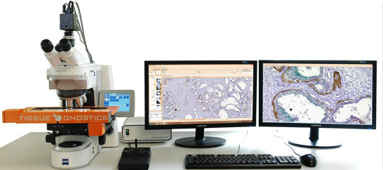
The cutting-edge tool for cell-based staining intensity analysis of immunohistochemical routine samples and research experiments.
The HistoFAXS analysis system puts all relevant parts for effective and precise cell and tissue analysis together. From scanning of brightfield samples to full and detailed digital image processing with analysis, HistoFAXS will satisfy the everyday requirements of laboratory workers. The combination of high-tech hardware from Zeiss, Leica, Nikon microscopes and fast multiple-core computer workstations with intuitive and easy-to understand software solutions bring your lab to the 21st century.
HistoFAXS is a microscopic system that automatically acquires immunohistochemically stained sections and performs quantitative analysis of staining intensities.
HistoFAXS is a easy to use, microscopic analysis system that automatically acquires up to 8 slides with immunohistochemically stained sections and performs quantitative analysis of staining intensities.
HistoFAXS unique features
- Automatic acquisition of an unlimited number of regions of interest on up to 200 slides
- Large overview images created from individual fields of view (FOV) may be exported at user-defined resolution
- Defines regions to be analyzed (or excluded from analysis) on the acquired regions of interest
- Automated, for two markers and Semi-automated for multiple markers, color separation to extract the relevant marker information out of immunohistochemical images
- Reliable automatic nuclear segmentation with minimum user interaction
- Forward Gating from the individual cell in the image to the dot in the scattergram
- Backward Gating from the individual dot or dot group to the corresponding cells in the sample
- DotPlot operations – analysis of the image processing results via gating and additional DotPlots
- Histograms and overlays
- Statistics including percentage of positive cells, cells/mm2, and mean intensity
New standard for the entire process of slide scanning and data handling
Cutting-edge tool for cell-based analysis of staining intensities of immunohistochemically stained sections
HistoFAXS is a high tech system offering automated routines for comprehensive procedures. Still, its handling is remarkably simple. The analytical process is based on standardized image acquisition. It is not linked to specific staining types or object sizes.
Automated workflow
HistoFAXS is a combination of high-end hardware modules (Zeiss, Leica or Nikon-based) and two software modules:
-
- HistoFAXS image acquisition and data management module.
- HistoQuest analysis module for immunohistochemical stainings (stand alone use possible).
HistoFAXS provides a smooth, automated workflow from image acquisition to publication quality, output of graphs and images, as well as customizable data export for further processing。
HistoFAXS Tissue Microarray
Tissue Microarray (TMA) module is fully integrated in the TissueFAXS system. Acquisition is handled in the HistoFAXS software while the analysis is done in the HistoQuest Analysis Software.
TMA Core detection
TMA spots are identified on a slide preview obtained by a low magnification scan. Acquisition can be made automatically, with manual adjustment or semi-manually by projecting TMA block patterns and doing comprehensive block operations. Each spot is given its individual ID based upon identification. Missing spots are still recognized based on block matrix.
TMA Groups
Logical groups can be drawn and named on the preview image, thus providing one possible basis for later analysis. After acquisition, the project is ready for analysis.
TMA Analysis
TMA projects are opened in HistoQuest. Logical groups are shown in HistoQuests Input Region list and are analyzed as one sample.
TMA spots not in logical groups are displayed separately in this list under their spot ID. They can be analyzed individually.
After analysis, results can be exported to Excel sheets to be linked with metadata for further examination.
A browser-type TMA explorer and a report generator completes the TMA functionalities.
