生物力学相关产品及科研服务
计算机控制的自动微吸管系统
型号:
联系人:李胜亮
联系电话:18618101725
品牌:匈牙利cellsorter
计算机控制的自动微吸管系统
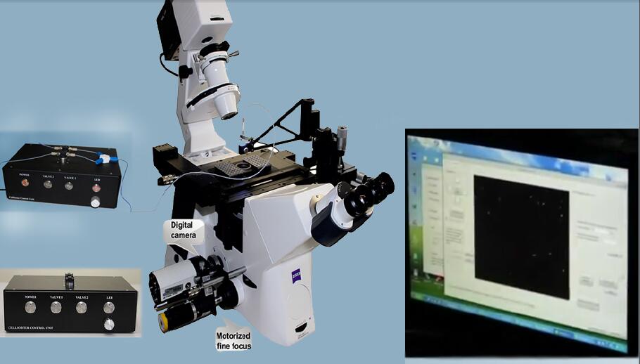
该系统是一种安装在正常倒置显微镜上的计算机控制的微量吸管系统,量化细胞形变、细胞间粘附作用和细胞与基质之间的粘附作用,可同步实现荧光观测、单细胞捕获、分选和分离和图像视频处理
一个可在已有显微镜上搭建的,可同步实验吸吮加载、图像视频处理与荧光观测的细胞力学装置。可建立微管吸吮力学加载-荧光观测耦合的分子-细胞动力学实时原位观测系统。
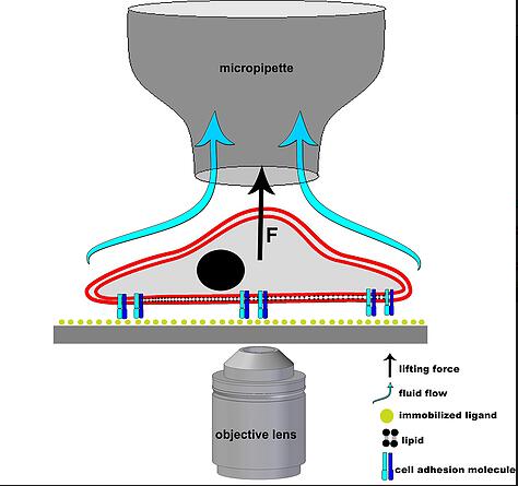
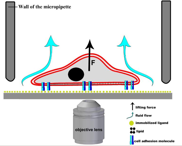
单个细胞粘附力测定模式图:
细胞与细胞之间粘附力测定模式图:
通过施加负压将细胞的一部分(或整体)吸入微管中,测量一定负压吸吮作用下细胞的变形及其时间历程或细胞粘附分离的临界负压来评价细胞的变形特性,同时记录细胞的吸入量。
图像处理技术和力学模型,量化测量细胞形变、细胞对之间的相互作用以及黏附特性、基质附着细胞测定细胞硬度等力学特性。
压电微管吸吮模块:对样品池内细胞的捕获、分选、吸吮和微操控;
集成倒置荧光相差显微镜:用于对所述细胞进行荧光激发;
信号采集模块:用于对微弱荧光信号的采集;
控制模块:用于对微管吸吮和荧光采集同步触发,并进行数据分析处理
特点:
-
1)High throughput single cell sorting directly from the Petri dish
-
One single cell arrives to each PCR tube
-
10 PCR strips containing 80 tubes can be filled in a cycle
-
Glass cover slip for testing single cell deposition in situ
-
Drop volume less than 1 ul for adherent cells
-
Pick up volume of ~1 nl for suspended cells
-
15-20 seconds per cell. When collecting multiple cells, sorting speed is 1 cell/second.
-
Number of cells picked up in a single run: 1-1000.
-
Isolates a subpopulation of live adherent cells expressing fluorescent or luminescent markers
-
Both unlabeled and fluorescentcells are recognized by computer vision
-
Viable cells after sorting
-
Any adherent and non-adherent cell type can be sorted
-
Cell culture needs minimal preparation before sorting
-
Average sorting process takes only a few minutes
-
Multichannel detection using the fluorescent filter setup of the microscope
应用范围:
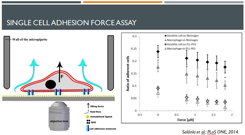
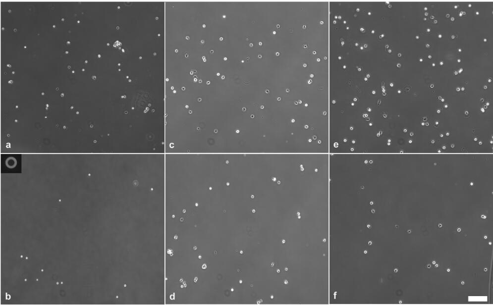
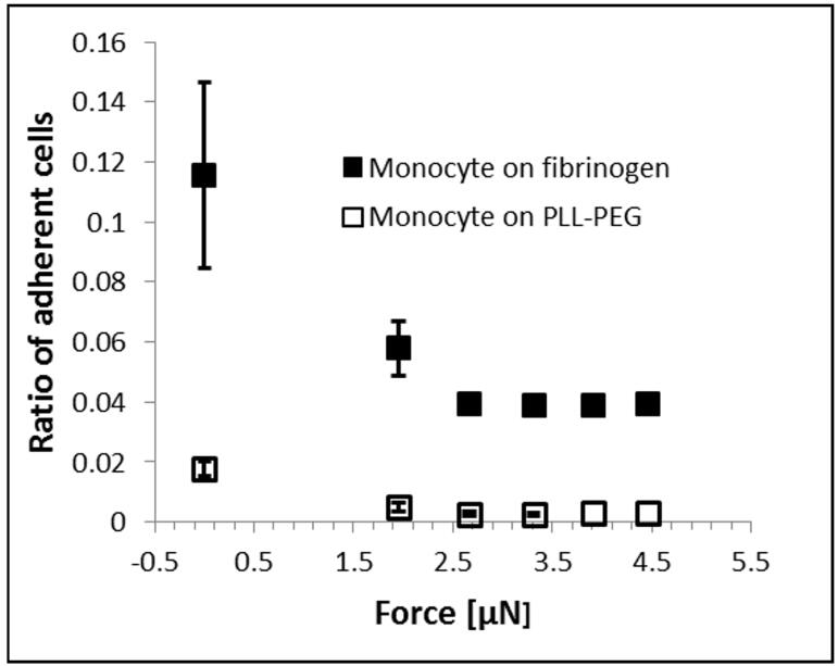
1)计算机控制的自动化微吸管系统测定单细胞黏附力(Single Cell Adhesion Assay Using Computer Controlled Micropipette)
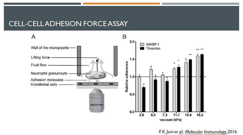
2)计算机控制的自动化微吸管系统测定细胞与细胞之间的黏附力(Cell-cell adhesion force assay Using Computer Controlled Micropipette)
-
R. Salánki et al. : Single cell adhesion assay using computer controlled micropipette, PLoS ONE 9(10): e111450 (2014) Open access paper.
-
P. K. Jani et al.: Complement MASP-1 enhances adhesion between endothelial cells and neutrophils by up-regulating E-selectin expression, Molecular Immunology 75, 38–47 (2016)
-
N. Sándor et al.: CD11c/CD18 Dominates Adhesion of Human Monocytes, Macrophages and Dendritic Cells over CD11b/CD18, PLoS ONE 11(9), e0163120 (2016) Open access paper.
其他应用:
3)单细胞分离与抓取分选系统 (Single Cell Isolation and Sorting)
-
Z. K?rnyei et al.: Cell sorting in a Petri dish controlled by computer vision Nature Scientific Reports 3, Article number: 1088 (2013) Open access paper.
-
R. Salánki et al.: Automated single cell sorting and deposition in submicroliter drops, Appl. Phys. Lett. 105, 083703 (2014)
-
R. Salánki et al.: High-throughput image based single cell isolation, Microscopy and Analysis, January issue, S10-13 (2015) Open access paper.
-
R. Ungai-Salánki et al.: Automated single cell isolation from suspension with computer vision, Scientific Reports 6, Article number: 20375 (2016) Open access paper.
4)循环肿瘤细胞(CTC)分离 (Circulating tumor cell (CTC) isolation)
Marnie Winter et al.: Isolation of Circulating Fetal Trophoblasts Using Inertial Microfluidics for Noninvasive Prenatal Testing, Advanced Materials Technologies 1800066 (2018)
5)单细胞RNA测序 (Single Cell RNA Sequencing)应用文献
-
Mia Palmkvist: Malaria and polypeptides of plasmodium falciparum at the infected erythrocyte surface, PhD Thesis, Karolinska Institutet, Stockholm (2016)
-
A. Kozlov et al.: A screening of UNF targets identifies Rnb, a novel regulator of Drosophila circadian rhythms, The Journal of Neuroscience 7, 3286-16 (2017)
-
M. Ngara et al.: Exploring parasite heterogeneity using single-cell RNA-seq reveals a gene signature among sexual stage Plasmodium falciparum parasites, Experimental Cell Research (2018)
6)蛋白工程(Potein Engineering)
-
K. Piatkevich et al. : A robotic multidimensional directed evolution approach applied to fluorescent voltage reporters, Nature Chem Biol, doi:10.1038/s41589-018-0004-9 (2018)
