VesselMap血管分析软件

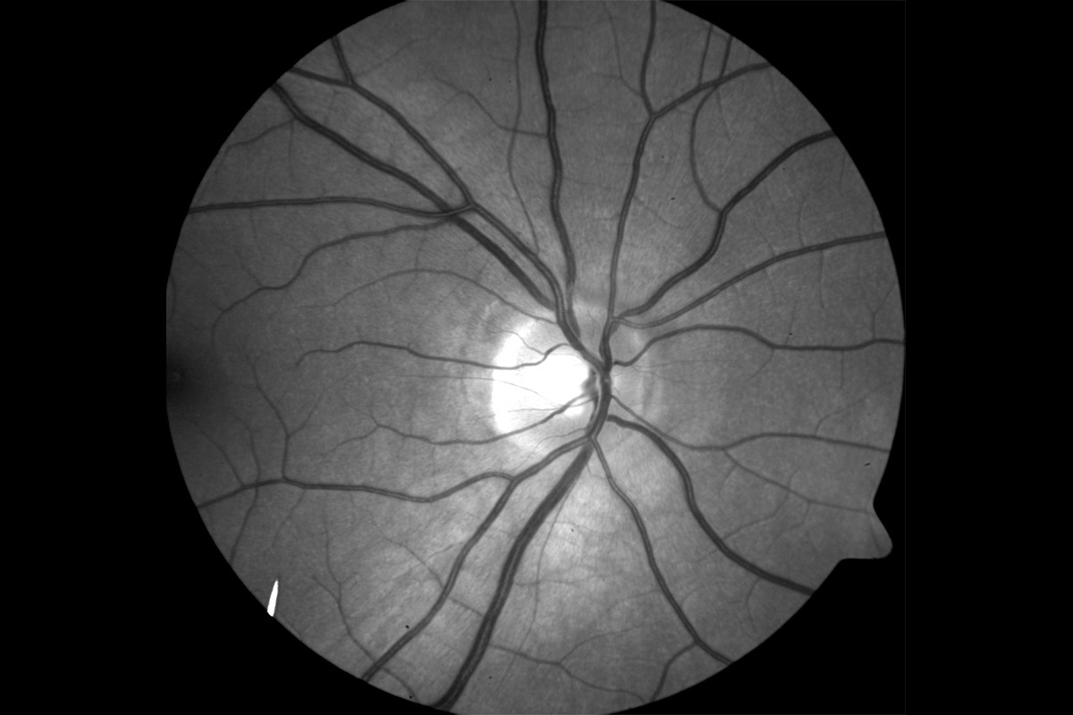
 VesselMap aric: Image of the ocular fundus
Analysis Software VesselMap
我们创新的分析软件?VesselMap aric?可以进行静态血管分析,以评估和评估视网膜血管的状况。它是我们静态血管分析系统的标准组件。
Our innovative analysis software ?VesselMap aric? enables static vessel analysis in order to assess and evaluate the condon of retinal vessels. It is available as a standard component of our systems for static vessel analysis.
Features & Benefits
Simple, programme-guided application
Semi-automatic and precise determination of vascular parameters
Suitable as screening method and for use in individualized medicine
Integrated function for follow-up examinations to significantly improve reproducibility and fully automatic evaluation of subsequent images
Patient-related storage of results
When used in combination with a non-mydriatic imaging system, no dilation of the pupil is required for the examination
Testing principle
Images from the ocular fundus are taken by using an imaging system.
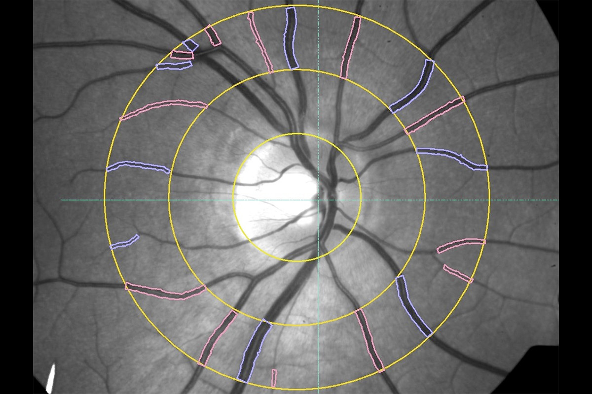
The fundus image is opened in the software and the papilla is marked. Therefore, a measurement grid is placed on the image.
Subsequently, within this measurement grid (ring zone), all essential arterial and venous vessels are marked manually by selecting and clicking on them.
The software now determines the vessel diameters according to the marked vessels and calculates the static vessel parameters.
The results are presented in a protocol.
The static vessel parameters include:
Central retinal arteriolar equivalent (CRAE): arterial model vessel diameter
Central retinal venular equivalent (CRVE): venous model vessel diameter
Arteriolar-to-venular ratio (AVR): CRAE/CRVE ratio
Using the follow-up function, steps 2, 3 and 4 are performed automatically.
The recording of the fundus images, determination of the parameters and evaluation of the static vessel parameters are carried out on the basis of the ARIC study.
The vascular parameters are valid biomarkers of the retina and describe the condon of the small arteries and veins of the retinal microcirculation. They can be used as risk factors or prognosis indicators for vascular diseases and events in the eye as well as other organs.
The AVR provides important information on cardiovascular risk. Changes in vascular parameters that occur between the individual examinations provide information on the progression of diseases and therapeutic effects.
Customised solutions – easy integration into existing systems
The analysis software VesselMap aric can also be used with existing imaging systems (fundus camera) and hardware components. For this purpose, the analysis and calculation parameters of the software are adapted to your system and adjusted accordingly. Individual solutions offer you addonal benefits, including:
Protocol functions individually tailored to your practice
Doctor-specific patient database for analysis results
Direct network integration
Image transfer with different image standards such as Dicom
In order to combine the software with your existing imaging system, the following hardware requirements must be met:
Suitable fundus camera
Laptop or PC (Windows 10)
Our Customer Service is happy to advise you and check if your existing hardware is compatible with our products. Contact us for more information!!
Addonal modules
We offer the following modules for addonal application areas:
Research Option: For freely selecting vessel sections
Contact us for more information!!
