 Abstract
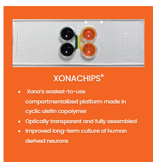
Xona Microfluidics, Inc. has developed a new easy-to-use microfluidic chip to compartmentalize neurons and establish microenvironments, called the XonaChips?. XonaChips? are made out of an optically transparent plastic, cyclic olefin copolymer, which allows us to offer fully assembled chips that require few preparation steps, and result in excellent neuronal growth. Importantly, XonaChips? are compatible with high-resolution fluorescence imaging.
Download Post as PDF
Introduction
Traditional neuron culture approaches result in random outgrowth of axons and dendrites, which prevent the study of neurons in their unique polarized morphology. Microfabricated multicompartment devices, pioneered by Xona scientists, have become well-established and well-used research tools for neuroscientists in the last 10-15 years (selected high profile publications are referenced 1-17). These devices compartmentalize neurons and provide a method to physically and chemically manipulate subcellular regions of neurons, including somata, dendrites, axons, and synapses 19-20. They also provide multiple experimental paradigms that are not possible using random cultures, including studies of axonal transport, axonal protein synthesis, axon injury/regeneration, and axon-to-soma signaling.

To provide an easy-to-use and fully assembled device, Xona has now developed plastic XonaChips? (Fig. 1)18. The interior of the XonaChips? are made permanently hydrophilic, simplifying device wetting. Further, these chips are fabricated in an optically transparent plastic, cyclic olefin copolymer, ideally suited for high resolution imaging.
Methods
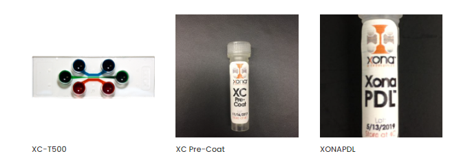
XonaChips? were prepared according to the protocol. XC Pre-CoatTM was first used to ensure even wetting of the chip. XC Pre-CoatTM is included with each XonaChips?order. The chip was then coated with XonaPDLTM, which includes the optimal concentration of poly-D-lysine for Xona’s devices. The chip was washed multiple times with PBS and then filled with neuronal culture media.
E18 Hippocampal Rat Neurons were dissociated and prepared for loading into the XonaChips? as described in the protocol. Approximately 120,000 neurons were seeded into the right compartment of the XonaChips?. Images were acquired with a laser scanning confocal microscope using a 30x/1.05 N.A. silicone oil (ne = 1.406) objective lens.
Results & Discussion
To evaluate the performance of the XonaChips?, researchers at the University of Carolina at Chapel Hill grew rat hippocampal neurons within the chip for 21 days. As illustrated, axons are healthy and isolated within the axonal compartment (Fig. 2).
To demonstrate the ability to create isolated microenvironments, researchers added a low molecular weight fluorescent dye (Alexa Fluor 488 hydrazide) to the axonal compartment. The dye stayed isolated to the axonal compartment consistent with Xona’s silicone devices17.

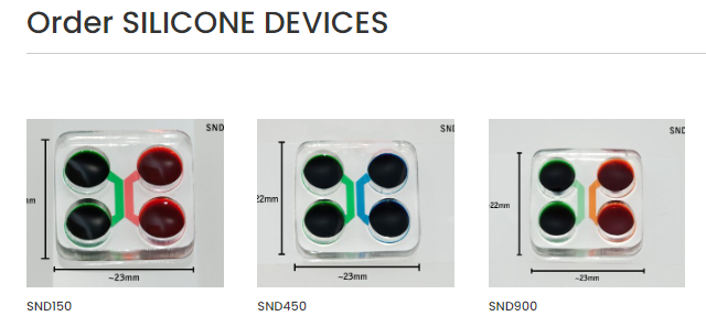
Researchers at UNC-Chapel Hill also evaluated high-resolution live imaging within the XonaChip?. The XonaChip? consists of a molded COC top bonded to a thin optically transparent 150 ?m thick COC film. All excitation wavelengths tested (405, 488, 568, 647 nm) resulted in comparable results to images acquired in SND series devices attached to glass coverslips (data not shown). Because of the high quality of COC production, there is no autofluorescence of the material. Fig. 2 demonstrates the high image quality. A variety of objectives have been tested from 10x to 60x silicone oil. In all cases, results were comparable to glass coverslips.
Conclusion
In summary, the XonaChip? is a fully assembled multicompartment device that is easy to use and results in healthy, long-lasting cultures. These chips allow microenvironments to be established as with our SND series devices and are equally compatible with high-resolution fluorescence microscopy.
About Xona Microfluidics, Inc.
Xona Microfluidics, Inc. is a North Carolina corporation located in Research Triangle Park, North Carolina. More information can be found at xonamicrofluidics.com
