数字图像相关(Digital Image Correlation,DIC)也就是数字图像相关方法是一种非接触式的高精度位移、应变测量方法,是目前实验力学领域内有应用前景的测量方法。
测量quan场应变广泛应用于组织材料力学、断裂力学、微观纳米应变测量、各种新型材料测量等。该测量具有非接触性、应用广泛、精度较高、quan场测量、 数据采集简单、测量环境要求不高、易于实现自动化等优点,可以测量微米甚至纳米的变形。
是一种对材料或者结构表面在外载荷或其他因素作用下进行场位移和应变分析的新的实验力学方法。目前DIC技术已经在电子封装、材料测试、断裂力学、航空航天、生物力学以及显微测量等众多领域得到应用,取得了瞩目的成就
数字图像相关性(DIC)是一种光学方法,它使用跟踪和图像配准技术对样品在变形过程中的变化进行j确的2D和3D测量。 这通常用于测量变形过程中斑点图案样本的变形,位移,应变和光流。
Articulation-Induced Responses of Superficial Zone Chondrocytes in Human Knee Articular Cartilage - Effects of Shear and Sliding
Introduction: During daily physical activities, joint articulation results in 3-10% compression in its overall thickness. There is also consensus that articulation is a combined process of shearing and sliding, with relative rotational and translational motions between femoral condyle and tibia cartilage at least at the millimeter scale 2,3. The macroscale motions in the joint which translates to microscale tissue deformation have been assessed in vitro to range from 2.8 to 41.0% depending on the tissue state and lubrication 4 . Biologically, dynamic shear at small amplitudes (±3%) markedly stimulates PRG4 secretion by immature bovine cartilage disks, but little evidence has been provided for similar effects in human cartilage, perhaps due to insufficiently large amplitudes of stimulation 5 . A prior study on the effects of torsional shear on chondrocyte apoptosis has suggested that superficial zone (SZ) cell apoptosis occurs in bovine explants when sheared without lubrication 6 . However, it is unknown whether (1) the degree of SZ cell death and amount of PRG4 secretion is a function of shear and sliding amplitudes and (2) how much resultant tissue deformation and sliding occurs during the chosen method of shear. Therefore, the hypothesis for this study was that articulation induces an amplitude-dependent effect on cartilage superficial zone health and responses, and that the variations in responses are due in part to the extent of tissue shear. Thus, the aims were to assess the effects of graded levels of articulation on chondrocyte viability and PRG4 secretion, and to relate these biological responses to the level of tissue shear.
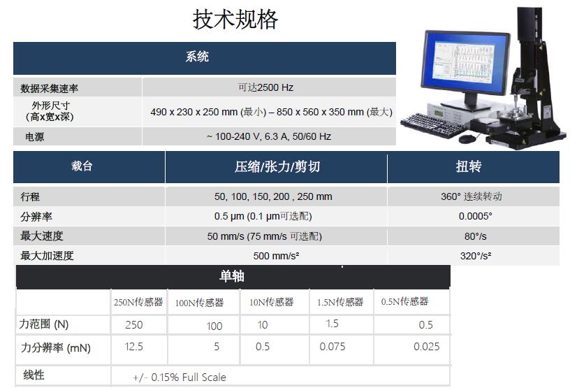
Methods: A total of 25 human articular cartilage disks (2mm diameter, 1.1mm thick) were harvested from the patellofemoral groove of each of 9 cadaveric knees of donors spanning a wide age range [n=3 Young (<30 yrs), 19, 26 and 29 yrs; n=6 Old (>50yrs), 51, 54, 55, 61, 64, and 66 yrs] without evidence of osteoarthritis; the samples were prepared to include the articular surface, and from regions of tissue with a macroscopically intact articular surface. Disks were incubated in medium supplemented with 10% FBS and 25 ?g/ml ascorbate. On day 3-5, some disks were subjected to mechanical articulation at Low, Mid, or High amplitudes, and others left free-swelling (None). Immediately after treatment, some disks were analyzed for chondrocyte viability at the articular surface. Other disks were incubated for two additional days to assess effects of Low and High articulation on subsequent PRG4 secretion. Some of the donor-matched disks were analyzed mechanically to assess the effects of Mid and High articulation on shear strain and shear stress. Mechanical stimulation. Articulation was implemented at sliding amplitudes of ±100?m, ±500?m, or ±1000?m using a sinusoidal 1Hz waveform in order to simulate effects of Low, Mid, or High levels, respectively. The articulation was imposed for 400 cycles and superimposed upon 20% static compression using a MachOne mechanical tester (BMM ) with a surface-finished polysulfone platen for articulation against the articular surface 7 and a roughened surface and trough to stabilize the deep cartilage surface. Multi-axial load was recorded. Other disks were maintained free-swelling (None) to serve as control. SZ cell viability analysis. Disks were stained with Live/Dead® (Invitrogen) immediately after treatment, and en face images of the disks were taken at 10x magnification with a fluorescent microscope. The resulting live and dead images were analyzed using a custom image-processing program to determine the number and percentages of live and dead cells. PRG4 protein secretion.. Conditioned medium, collected during the two days of incubation after articulation, was analyzed for PRG4 by ELISA. Biomechanical analysis of tissue shear. The response of cartilage disks was then assessed by video analysis. Videos were recorded with a Nikon D90 SLR digital camera, fitted with a macro lens, and frames analyzed for disk and platen position to determine shear deformation during imposed articulation. Statistics. Summary statistics are presented as mean ± SEM, with n = # of cartilage disks per experimental condition per donor knee, and m = # of donors, so that n*m = total cartilage disks per condition. Data that ranged over an order of magnitude were log10 (1+Y) transformed and percentage data were arc sine transformed prior to statistical analyses to improve normality and homoscedasticity. The effects of mechanical treatment on SZ cell death was assessed by 1-way ANOVA and post-hoc Tukey test. Linear regression of both maximum shear stress and strain to cell death were performed. The effects of age and mechanical stimulus on cartilage PRG4 secretion were assessed by 2-way ANOVA with donor as a random factor.
Results: Articulation induced chondrocyte death in the superficial zone in an amplitude-dependent manner (Fig. 1). In control samples and those subjected to Low articulation, SZ cell viability was high at 90-95%. Mid and high amplitudes of applied articulation resulted in a reduction of viability by 20-25% (p<0.01). Cartilage PRG4 secretion was modulated by age group (p <0.001) and articulation stimulus (p<0.001) in an interactive manner (p<0.01, Fig. 2). In the absence of applied mechanical stimulation, PRG4 secretion by cartilage from young donors was higher (+4.7 ?g/(cm2?day), p<0.05) than that of old donors. With mechanical stimulation, higher articulation resulted in higher PRG4 secretion (+4.0 and +6.8 ?g/(cm2?day), respectively, for low and high articulation) in young donors, but did not have a discernible effect on cartilage from old donors (-0.04 and -0.4 ?g/(cm2?day), for low and high shear, respectively). Cell death and cartilage PRG4 secretion responses were associated with donor-specific tissue mechanics ( Fig. 3 ). Positively correlations were found between cell death and shear stress (Fig. 3A & C, p<0.05) as well as PRG4 secretion and shear strain (p<0.05). Trends were observed between cell death and high shear strain, but were not significant (Fig.3B, p=0.10).
Discussion: The finding that high levels of cartilage shear stress and/or strain can lead to cell death suggests the susceptibility of cartilage to states when lubrication is deficient and/or tissue asperities lead to high articulation-induced tissue shear. On the other hand, the stimulated PRG4 secretion response suggests that the remaining cells can exhibit a feedback response, attempting to counter the excessive shear, as suggested previously 5 . The correlation of biological effects with tissue-specific biomechanical properties suggests that age-related variations in human articular cartilage properties could be involved in variations in mechanobiology. The lack of mechanical responsiveness of human articular cartilage with aging may contribute to the increased incidence of osteoarthritis with aging.
Significance: The susceptibility of articular cartilage to shear-induced cell death suggests that mechanical protection strategies may be helpful to prevent subsequent development of osteoarthritis. Further understanding of the mechanobiological alteration of human cartilage with aging may involve both mechanical properties of the tissue matrix as well as biological variations in the chondrocytes.


